 |
NUCLEAR MEDICINE
What is General Nuclear Medicine? Nuclear medicine is a subspecialty within the field of radiology. It comprises diagnostic examinations that result in images of body anatomy and function. The images are developed based on the detection of energy emitted from a radioactive substance given to the patient, either intravenously or by mouth. Generally, radiation to the patient is similar to that resulting from standard x-ray examinations. What are some common uses of the procedure? Nuclear medicine images can assist the physician in diagnosing diseases. Tumors, infection and other disorders can be detected by evaluating organ function. Specifically, nuclear medicine can be used to:
How should I prepare for the procedure? Usually, no special preparation is needed for a nuclear medicine examination. However, if the procedure involves evaluation of the stomach, you may have to skip a meal before the test. If the procedure involves evaluation of the kidneys, you may need to drink plenty of water before the test. How does the procedure work? You are given a small dose of radioactive material, usually intravenously but sometimes orally, that localizes in specific body organ systems. This compound, called a radiopharmaceutical agent or tracer, eventually collects in the organ and gives off energy as gamma rays. The gamma camera detects the rays and works with a computer to produce images and measurements of organs and tissues. How is the procedure performed? A radiopharmaceutical agent is usually administered into a vein. Depending on which type of scan is being performed, the imaging will be done either immediately, a few hours later, or even several days after the injection. Imaging time varies, generally ranging from 20 to 45 minutes. The radiopharmaceutical that is used is determined by what part of the body is under study, since some compounds collect in specific organs better than others. Depending on the type of scan, it may take several seconds to several days for the substance to travel through the body and accumulate in the organ under study, thus the wide range in scanning times. While the images are being obtained, you must remain as still as possible. This is especially true when a series of images is obtained to show how an organ functions over time. After the procedure, the physician will carefully review the images and send a report to your doctor. What are the benefits vs. risks? Benefits
Risks
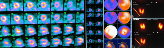
Cardiac Nuclear MedicineWhat is Cardiac Nuclear Medicine? Cardiac nuclear medicine refers to these diagnostic tests that are used to examine the anatomy and function of the heart. Cardiac nuclear medicine tests are indicated for individuals with unexplained chest pain or chest pain brought on by exercise (called angina) to permit the early detection of heart disease. The most common cardiac nuclear medicine procedure, called myocardial perfusion scanning, enables the visualization of blood-flow patterns to the heart walls. The test is important for evaluating the presence and extent of suspected or known coronary artery disease (blockages) as well as the results of previous injury to the heart from a heart attack, called a myocardial infarction. It also can be done to evaluate the results of bypass surgery or other percutaneous revascularization procedures designed to restore the blood supply to the heart. Heart-wall movement and overall heart function can be evaluated with cardiac gating, a technique that synchronizes the images of the heart with different parts of the cardiac cycle (contracting or relaxing) as determined by an electrocardiogram (ECG), which records the electrical currents that activate the heart muscle and cause it to pump. How should I prepare for the procedure? You should avoid caffeine (coffee, tea, etc.) and smoking for 48 hours before the examination. You should not eat or drink anything after midnight before the procedure, but continue taking medications with small amounts of water unless your physician says otherwise. Wear comfortable, walking shoes and loose-fitting clothes for your procedure. Tell the technologist and supervising physician if you have asthma or a chronic lung disease or have problems with your knees, hips or keeping your balance, which may limit your ability to perform the exercise needed for this procedure. How does the procedure work? Coronary arteries are best evaluated by determining the changes in blood flow to the heart due to exercising. Consequently, you will undergo a stress test-most commonly through physical exercise-to make your heart work harder than normal. Then you will be given a radioactive compound, called a radiopharmaceutical agent or tracer. This compound will collect in parts of your heart with good blood flow and will give off gamma rays. The gamma camera detects the rays. Subsequently, a computer following a set of complicated mathematical formulas will construct images of the heart based on the detected gamma rays. How is the procedure performed? For the stress part of the examination, you will exercise by either walking on a treadmill or pedaling a stationary bicycle for a few minutes. While you exercise, the electrical activity of your heart will be monitored by electrocardiography (ECG), and your blood pressure will be measured frequently. Before you stop exercising, you will be given the radiopharmaceutical through an IV leading into a vein in your arm. The compound is given when the blood flow to the heart is at its peak because of your exercising. This provides the best opportunity to identify regions of the heart that are not receiving adequate blood flow. One minute later, you will stop exercising. Approximately one half-hour later, as you lay on an examining table, the compound will have collected in your heart. The gamma camera will then be used to obtain images. The gamma camera likely will move slowly and automatically in an arc over the front of your chest after it is positioned initially by the technologist. The images obtained after exercise must usually be compared with images of your heart obtained after injection of the same radiopharmaceutical while you are resting. This may be performed before or after the exercise part of the examination, depending on the protocol used. Comparison of the exercise and resting images is done to determine whether coronary blood flow has changed once you have rested and to check for coronary artery disease. If you are unable to use a treadmill or bicycle, you will not exercise, but you will be given a drug that will cause your heart to work as hard as if you had exercised. You will then be given the radiopharmaceutical. Immediately after the procedure, a diagnostic radiologist with specialized training in nuclear medicine will check the quality of the images to ensure that an optimal diagnostic study has been performed. What will I experience during the procedure? You may experience some minor discomfort from the intravenous injection of the radiopharmaceutical. You will be asked to exercise until you are either too tired to continue or short of breath, or if you experience chest pain, leg pain, or other discomfort that causes you to want to stop. If you are given a medication to increase blood flow because you are unable to exercise, the medication may induce a brief period of feeling anxious, dizzy, nauseous, shaky or short of breath. In rare instances, if the side effects of the medication are severe or make you too uncomfortable, other drugs can be given to stop the effects. Most patients can resume regular activities immediately after the procedure. The radioactivity in your body will decrease due to the natural process of radioactive decay. In addition, radioactivity will decrease because the radiopharmaceutical passes out of the body in the urine or stool. What are the benefits vs. risks? Benefits
Risks
Source: RadiologyInfo |
|

|